Hindfoot Arthritis
What is the Hindfoot?
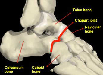
The hindfoot generally refers to the section of the foot that begins immediately below the ankle joint and ends at the level of the Chopart joint. You cannot feel the Chopart joint but it is made up of 2 joints in line, called the talonavicular joint and the calceneocuboid joint and is found below and in front of the ankle joint.
What bones are contained within the Hindfoot?
The hindfoot contains 2 main bones called the talus and the calcaneum (heel bone) that are connected by “joints” to one another and also these bones are connected in front to the “midfoot” area where the hindfoot meets the navicular and cuboid bones at the “Chopart joint”.
What causes Hindfoot arthritis?
 There are different reasons for developing arthritis in the hindfoot:
- Post-traumatic
Bone fracture, even if successfully treated many years before, can predispose the area to developing arthritis.
- Rheumatoid arthritis
Patients with rheumatoid or other forms of inflammatory arthritis can develop arthritis in their hindfoot
- Osteoarthritis
Even in the absence of injury, arthritis can develop unexplained in the hindfoot
- Tibialis posterior tendon dysfunction
If this tendon in the foot develops problems then the arch can flatten and arthritis can develop in the midfoot and hindfoot
What are the symptoms?
- Aching in the middle or back of the foot when walking/activities
- Loss of flexibilty in the foot, especially on uneven surfaces
- Swelling may become apparent around the ankle area and side of the foot
- The foot can subtly change its shape and become flatter in its appearance with loss of the natural arch, and the heel bone can begin to point outwards
- Wearing certain shoes can rub on the skin on the inside of the foot and the shoes may wear out quicker.
How is the condition diagnosed?
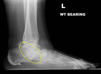 The clinical features as discussed above in addition to typical X-Ray findings seen can confirm the diagnosis. Further scans are usually unnecessary, but some surgeons will arrange a CT scan to exactly identify the joints involved.
Can the problem get worse?
People often “live” with the symptoms for many years and then finally seek medical attention. If you decide not to see your doctor, the problem will tend to progress, usually slowly. Stiffness will usually increase and the pain can get worse making walking gradually more difficult and weight bearing exercise difficult. The condition can be treated at any stage but as it develops, the hindfoot can slowly move out of shape and affect other nearby joints with arthritis.
How do you treat Hindfoot arthritis?
The treatment can involve a combination of different therapies below:
- Pain-killers and anti-inflammatories
- Shoe modification, often stiffer soled shoes or “rocker bottom” shoes can really help when walking, talking the strain off the painful joints
- Insoles moulded to the shape of the foot can support or correct deformity
- Lifestyle advice and avoidance of pain exacerbating activities
- Steroid/Anaesthetic injections into the joints under Ultrasound control, excellent at temporary relief (up to 6 months) and can be repeated
- Physiotherapy to keep your calf muscles relaxed and foot muscles strong can help
- Surgery
Surgery usually necessitates formal stiffening procedures of the arthritic joints involved. Depending on how many joints are affected one or all (there are 3) of the main joints can be part of a hindfoot fusion. This is a successful operation because the arthritic joints are already usually stiff and “painful” but following surgery although still stiff, they will be “painless”.
Deciding whether surgery is necessary
Many patients are simply seeking advice on managing a problem. In mild cases modifying shoes or activities can prove a successful therapy however if non-operative measures have failed to improve symptoms then surgery is indicated, but the timing of the surgery can be arranged to suit your needs. As said earlier, even if the condition worsens, surgery is always an option, but the exact surgical procedure performed may differ slightly from the original surgery possible and may be more technically challenging.